

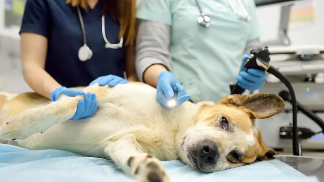
A veterinary endoscopy system is more than a scope. It combines access, visualization, documentation, accessories, reprocessing and support. The right configuration should make the clinical pathway clearer, not more complicated.
Start with the clinical pathways
Begin with the examinations and procedures your team performs today, then add the pathways you realistically plan to introduce. Upper gastrointestinal, ENT, airway, urinary and minimally invasive procedures can require different scope geometries and supporting equipment.
Patient size and species matter as much as procedure type. A system intended for small-animal ENT work will be configured differently from one designed for equine gastroscopy or a broad referral caseload.
Decide between portability and a workstation
Portable systems integrate the display and core controls into a compact host. They can suit focused examinations, point-of-care use and movement between rooms. A modular workstation provides more space for processors, displays, pumps, instruments, storage and documentation.
Consider where the system will be used, who will operate it, how often it will move and whether the practice needs one focused pathway or support for several scope families.
Compare the specifications that affect access
Specifications become useful only when connected to anatomy and workflow. Compare outer diameter, working length, channel size, angulation and optical geometry together.
| Specification | Why it matters | Confirm |
|---|---|---|
| Outer diameter | Access and patient suitability | Species, size and anatomy |
| Working length | Reach to target anatomy | Required examination pathway |
| Working channel | Suction, irrigation and instruments | Compatible tools and sampling needs |
| Angulation | Tip positioning and navigation | Direction and degree of bending |
| Field of view | Visible area and viewing angle | Optical geometry for the procedure |
Plan visualization and documentation
Review the complete imaging chain: processor or camera, light source, display, image and video capture, storage, output connections and case-management requirements.
Confirm how media is stored, whether reports are created within the platform and how records will be exported or archived. These details affect the workflow long after the examination itself.
Configure the complete system
A quotation may include only a scope, only the host or a complete system. Confirm the boundary between Included and Optional items for the scope, processor, display, trolley, pump, valves, instruments, cleaning accessories and training.
Compatibility should be confirmed as a system rather than one item at a time. This is especially important when several procedure types will share the same imaging platform.
Consider support across the equipment lifecycle
Review onboarding, instructions for use, reprocessing requirements, warranty, service routes, replacement parts and expected response times. Cleaning and reprocessing should follow the manufacturer’s current instructions and applicable protocols.
A practical decision checklist
- Define the procedures and patient range.
- Confirm scope diameter, length, channel and angulation.
- Choose between a portable host and a modular workstation.
- List the processor, display, pumps, instruments and accessories required.
- Confirm image capture, storage and report-output needs.
- Separate Included and Optional items in the quotation.
- Review reprocessing instructions, training, warranty and service support.
This article provides general equipment-planning information and is not a substitute for clinical judgment, product instructions for use or applicable reprocessing protocols.