

Veterinary Video
Otoscope
View the ear canal on screen and capture findings as you examine.
The LUMIVET video otoscope combines illumination, image capture and a 3-inch display in a handheld unit for veterinary ear visualization. For instrument use, select an external channel matched to the probe and instruments.


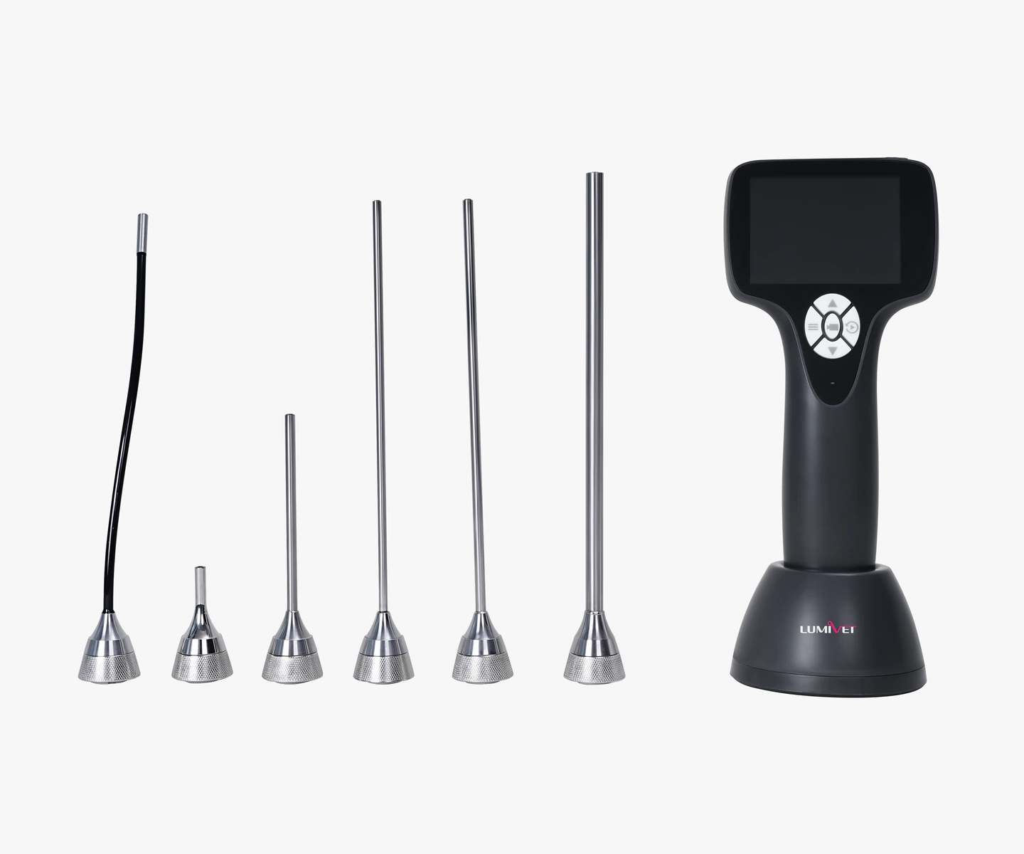
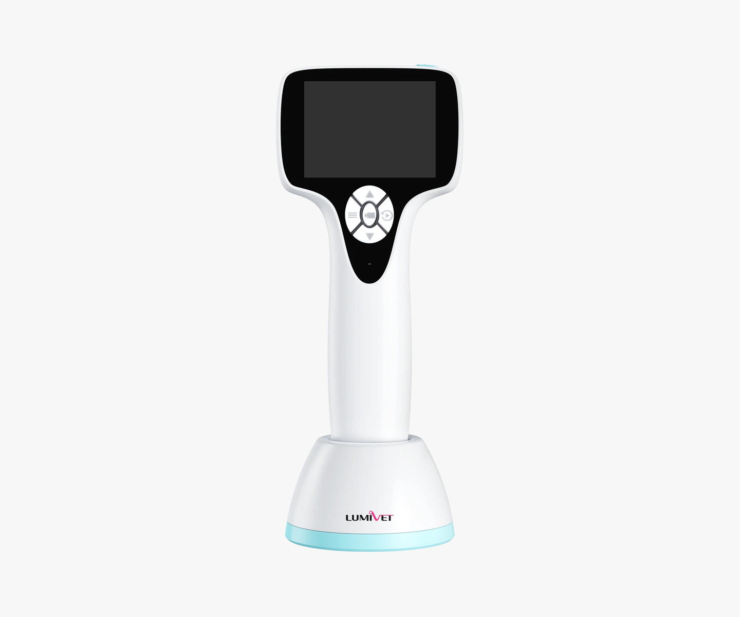
Configuration examples. Probes, interfaces and accessories depend on the selected setup.
View the examination on the handheld screen.
The live image appears directly on the unit’s 3-inch screen, with no external display needed for basic viewing. Veterinarians can watch the examination and show findings to colleagues or pet owners.

Capture findings for follow-up and communication.
Freeze images, take JPG photographs and record AVI video during the examination. Files are saved to a 64 GB Micro SD card, with playback, deletion and export available. Saved images can support later review, comparison over time and discussions with pet owners.
For case-based file management, use compatible computer workstation software. The handheld unit stores examination files but does not support case naming or provide a complete case-management system on its own.
Use compatible instruments through an external channel.
For sampling, irrigation or foreign-body handling under visual guidance, pair the probe with a compatible external channel and appropriate instruments. Match the channel, instruments and connection accessories to the planned task; supplied items are defined by the agreed configuration.
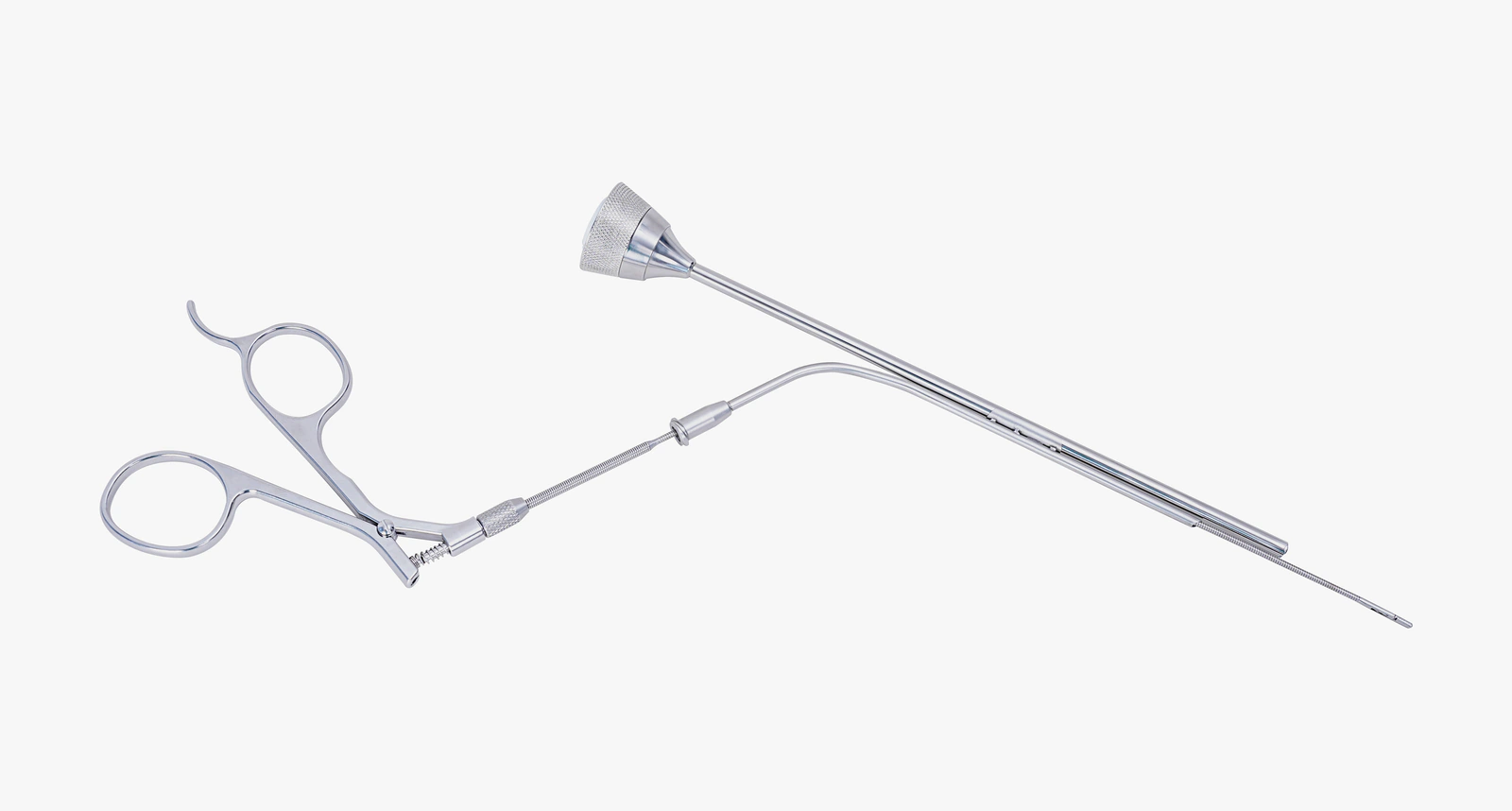
Select the probe and handheld unit for your examination.
Choose the probe for the intended viewing area and access conditions, then match the handheld unit’s interfaces, ear speculum, external channel and instruments. Additional probes are available in different diameters, working lengths and viewing directions. Check imaging specifications and accessory compatibility for each probe separately.
Standard ear probe
Reference specifications for the standard ear probe, order number 6100500334:
The standard ear probe has a maximum outer diameter of 4.2 mm, a working length of 30 mm, a 0° direction of view and a 120° field of view, with six white LEDs for illumination. The area that can be seen depends on access conditions and the probe and ear speculum configuration. A wide field of view does not ensure complete visualization of a curved ear canal.
Item
Standard ear probe reference value
Maximum outer diameter
4.2 mm
Working length
30 mm
Direction of view
0°
Field of view
120°
Camera resolution
1280×720
Illumination
Six white LEDs
The camera resolution above applies to the standard ear probe, not the handheld unit’s screen or all additional probes. The supplied model and accessories are defined by the agreed configuration list.
Handheld units and connections:
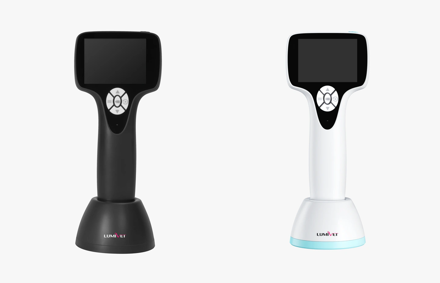
Item
Black unit
White unit
Built-in screen
3-inch LCD
3-inch LCD
Screen resolution
640×480 RGB
640×480 RGB
Storage
64 GB Micro SD
64 GB Micro SD
USB
Charging and photo/video file transfer
Charging and photo/video file transfer
mini HDMI
Connection to a compatible external display
Not provided
The dock provides a place to keep and charge the rechargeable handheld unit in the examination room. A USB charging cable is supplied as standard; a mini HDMI cable is optional. External display compatibility depends on the interface, signal and cable, not the connector’s physical shape alone.
The supplied handheld unit, probes, ear specula, dock, channels, instruments and other accessories are defined by the agreed configuration list.
Follow the handling instructions for each component.
The handheld unit, reusable probes and disposable ear specula have different handling requirements. Follow the instructions for the relevant model. Do not immerse the handheld unit; a probe’s protection rating does not apply to it. Ear specula are for single use; clean, disinfect and maintain reusable components according to the instructions for the selected configuration.
A one-year warranty is provided. Coverage and after-sales terms are defined by the supply agreement.
Confirm the otoscope setup for your examination.
Tell us the animal species, the area you intend to examine, and whether you need sampling, irrigation or foreign-body handling. We can review the probe, handheld unit interfaces, external channel and instrument combination accordingly. If you already have a display or workstation, include its model details so we can check connections and file-management options.