

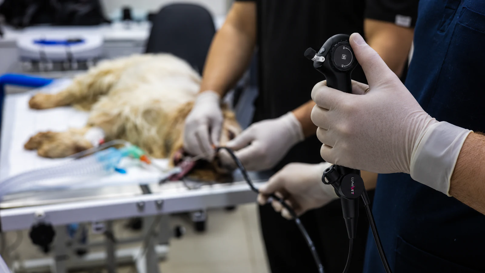
Composite illustration of a veterinary endoscopy setting using a representative LumiVET scope; not an actual clinical case.
Canine bronchoscopy allows a veterinary team to examine the inside of a dog’s trachea and bronchi. A flexible scope provides a direct airway view and access for selected sampling methods. Bronchoalveolar lavage (BAL) can provide samples from smaller airways and alveolar regions. These are complementary ways of investigating respiratory disease, not interchangeable tests. 1
For equipment planning, separate two questions: what does the team need to see, and what samples does it need to collect? The answers help define the scope, instruments, imaging equipment and supporting connections required for the examination.
What can bronchoscopy show?
A bronchoscope can reveal changes within accessible airways, including narrowing or collapse, abnormal tissue, secretions and foreign material. It may also allow retrieval of suitable airway foreign bodies when access, instruments and clinical conditions permit. 2
The visible airway appearance is only one part of the investigation. Cytology and microbiology can contribute information that an image alone cannot provide. Findings from samples must be interpreted in the context of the patient’s wider assessment. 3
For a practice comparing equipment, the distinction is important: a configuration intended for observation should not automatically be treated as a complete sampling or retrieval setup.
Applications described in veterinary literature
Veterinary literature describes bronchoscopy in the investigation of problems such as persistent cough, suspected airway foreign material, structural airway abnormalities, chronic bronchitis or pneumonia. Reported applications include examining airway structure, obtaining samples and supporting selected interventions. 2
These examples describe applications, not a checklist for deciding whether an individual patient should undergo bronchoscopy. That decision depends on the clinical question, patient assessment and relevant imaging. The extent of airway access also depends on the patient and the equipment used. 4
What does BAL add?
BAL is a sampling procedure in which sterile saline is introduced into a selected lung region and recovered for laboratory analysis. Depending on the clinical question, the fluid may be submitted for cytology, culture or other appropriate testing. BAL can be performed with bronchoscopic guidance or by a non-bronchoscopic technique in selected circumstances. 4
Bronchoscopic guidance combines airway inspection with sampling. Tracheal washes and BAL collect material from different parts of the respiratory tract, so they should not be described simply as better or worse versions of the same test. Selection depends on the information needed and the patient’s circumstances. 1
The distinction also explains why direct inspection and BAL may have different value in a case. An airway image does not directly examine the lung tissue beyond the visible airway surface. A sample can add cellular or microbiological information, but it does not replace the rest of the diagnostic work-up. 3
Equipment and team considerations
Respiratory patients require careful assessment of the risks associated with anesthesia, airway access and sampling. Equipment choice is part of that assessment, not a substitute for it. 4
Before selecting a configuration, the clinical and equipment teams should be clear about:
- The question the examination is intended to answer.
- Whether the plan is observation alone or includes sampling or retrieval.
- Which instruments and sample-collection accessories are required.
- Which existing imaging and recording equipment can be used.
- Whether the practice has the training, facilities and support needed for the planned work.
These questions can also support a referral discussion. They keep the choice focused on the intended examination rather than on purchasing the longest scope or the largest equipment package.
Choosing the scope and supporting equipment
Scope dimensions, instruments and imaging equipment need to be considered together. Veterinary endoscopy equipment varies in its intended applications and supporting components; a similar appearance does not establish compatibility. 5
Outer diameter and access
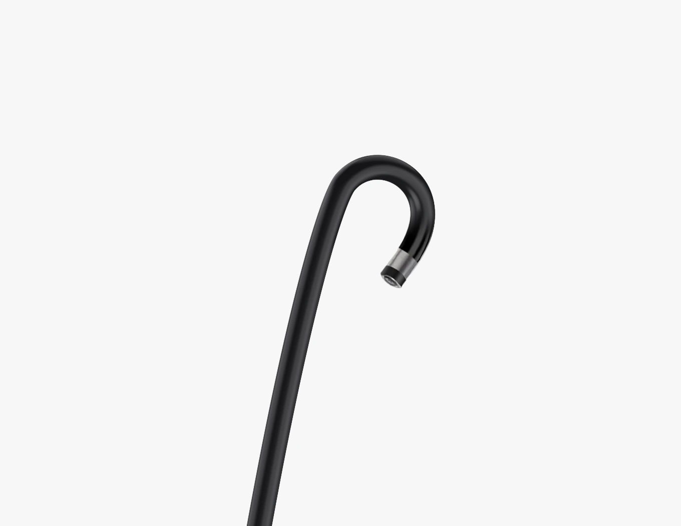
Review the animal’s actual airway dimensions and planned access route. Do not select a scope by bodyweight or breed label alone. A slim profile is one factor in the assessment, not a guarantee of suitability for every small patient.
Working length and tip control
Match working length to the intended examination. Confirm the directions and range of tip movement, along with how the operator will handle the scope and view the display. Extra length is not, by itself, a reason to prefer one configuration.
Channel and instrument fit
Check the scope’s channel inner diameter against the instrument outer diameter, and confirm sufficient instrument working length. Review the specified compatibility of each brush, forceps or other accessory rather than assuming that an instrument fits because it is labelled for endoscopy.
For lavage or suction, check more than the channel measurement. Connections, collection accessories and the suitability of the complete configuration require confirmation. A published channel diameter alone does not demonstrate that every proposed sampling method is supported.
Imaging and records
Confirm the compatible image-processing and display equipment. If still-image capture, video recording or export is needed, verify those functions on the selected equipment and software. They should not be assumed to be built into the scope itself.
Care and preparation
Include the manufacturer’s current instructions for use, component-specific cleaning or reprocessing requirements, and staff training in the equipment review. Do not apply a general endoscope cleaning method to every component. 5
LumiVET’s Veterinary Flexible Bronchoscope page lists the current reference dimensions and configurations. Use those details as a starting point for model-specific review, not as confirmation that every instrument or sampling accessory is compatible.
Keeping the examination record useful
Plan how images, observations and sample information will be brought together in the practice’s records. Useful equipment questions include whether the operator and assistant can share the view, how captured media is transferred, and how it is associated with the examination and laboratory results.
This is a workflow requirement to confirm with the selected system, rather than a reason to assume that every bronchoscope includes recording or case-management software.
Frequently asked questions
Are bronchoscopy and BAL the same procedure?
No. Bronchoscopy provides direct airway visualization; BAL is a method of collecting material from smaller airways and alveolar regions. They can be combined, but BAL can also be performed without a bronchoscope in selected circumstances. 1
Does a working channel mean the scope supports every sampling method?
No. The channel is only one part of compatibility. Instrument dimensions, connections, collection accessories, the manufacturer’s instructions and the intended clinical task all need review for the particular configuration.
Can one scope be selected for all dogs?
Suitability cannot be established by a single diameter or length specification. Patient anatomy, intended access, the examination plan and the complete equipment configuration need to be considered together.
Review the configuration for your practice
For airway equipment planning, review the Veterinary Flexible Bronchoscope configurations. Include the patients you treat, intended examination and sampling needs, and any existing imaging equipment when you contact LumiVET.
For a wider practice setup, explore the Specialty & Referral endoscopy options.
This overview is intended for veterinary professionals. It does not replace patient-specific clinical judgment, practical training or the instructions for the selected equipment.
References
- Finke MD. Transtracheal wash and bronchoalveolar lavage. Topics in Companion Animal Medicine. 2013;28(3):97–102. DOI: 10.1053/j.tcam.2013.06.003.
- Rha JY, Mahony O. Bronchoscopy in small animal medicine: indications, instrumentation, and techniques. Clinical Techniques in Small Animal Practice. 1999;14(4):207–212. DOI: 10.1016/S1096-2867(99)80012-7.
- Hawkins EC, DeNicola DB, Kuehn NF. Bronchoalveolar lavage in the evaluation of pulmonary disease in the dog and cat. State of the art. Journal of Veterinary Internal Medicine. 1990;4(5):267–274. DOI: 10.1111/j.1939-1676.1990.tb03120.x.
- Šmit I, Vučković M. Bronchoscopy and bronchoalveolar lavage in dogs and cats. Veterinarska stanica. 2024;55(4):471–484. DOI: 10.46419/vs.55.4.11.
- Callahan Clark J. Endoscopic Equipment for the Veterinary Practitioner. Today’s Veterinary Practice. November/December 2012.