

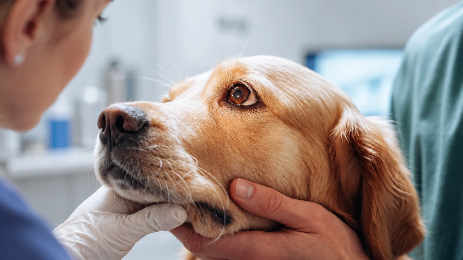
AI-edited illustration based on a licensed photograph, showing external nasal assessment—not rhinoscopy or an actual LumiVET clinical case.
Rhinoscopy in dogs provides an endoscopic view of the nasal passages and, when included in the examination, the nasopharynx behind the nasal cavity. It can help a veterinary team inspect abnormalities, obtain samples and retrieve suitable foreign material. The examination adds information to the wider workup; it does not provide a diagnosis in every case. 1
For equipment planning, distinguish what the team needs to see from what it needs to collect or retrieve. Observation, tissue sampling and foreign-body retrieval can require different scopes, instruments and supporting connections.
When veterinary teams consider rhinoscopy
Reasons to consider canine rhinoscopy include:
- Persistent nasal discharge, including discharge affecting one side.
- Epistaxis, or bleeding from the nose.
- Persistent sneezing or reverse sneezing.
- Suspected nasal obstruction, swelling or abnormal airflow.
- Suspected foreign material within the nasal passages. 1
These signs do not identify one particular disease. Timing and the choice of tests depend on the patient’s assessment, not a fixed requirement that every case first undergo prolonged treatment. The clinical team also considers whether the planned examination, anesthesia and sampling can be supported locally or require referral.
How rhinoscopy fits with imaging and sampling
Imaging and endoscopy answer different questions. CT can help assess the distribution of nasal disease, changes to bone, sinus involvement and dental pathology that may not be accessible to routine endoscopic inspection. The most appropriate imaging method depends on the clinical question; different modalities provide different information. 2
Rhinoscopy adds a view of accessible mucosal surfaces and an opportunity to obtain material for testing. A mass, inflammation or discharge seen on screen still needs interpretation alongside the examination, imaging and appropriate laboratory findings. Histopathology can be important, but one sample does not guarantee a definitive answer. 3
A useful equipment discussion therefore starts with the intended examination and sampling plan. A clear image and a suitable sample are related requirements, not the same requirement.
Anterior and posterior rhinoscopy
The two approaches assess different regions.
Anterior, or rostral, rhinoscopy examines accessible nasal passages through the nostrils. Posterior, or retrograde, rhinoscopy examines the choanae and nasopharynx from the oral side, commonly using a suitably flexible scope in a retroflexed position. An anterior view alone does not show all the same structures as a posterior view. 4
Whether both views are needed depends on the investigation. Scope selection needs to account for the different anatomy and maneuverability required by each approach.
What the examination involves
At an overview level, nasal endoscopy is performed under general anesthesia and may include visualization, clearing material from the field, sampling or selected foreign-body retrieval. 1
The team plans the examination alongside the patient’s assessment, imaging and sampling needs. Examination order also affects visibility: nasal instrumentation or sampling can introduce blood or debris that obscures later views. 4
Airway protection, positioning, instrument advancement and management of bleeding require clinical training and patient-specific decisions. They are not specified by an equipment overview.
What may be visible
Reported endoscopic findings include:
- Changes to mucosal color, surface or fragility.
- Excessive secretions or abnormal tissue.
- Narrowing, obstruction or altered visible space.
- Turbinate loss.
- Foreign material or plaque-like deposits. 4
These are observations, not final disease labels. Inflammation and tissue destruction, for example, can require further investigation rather than visual classification alone. In a recent study of canine nasal disease, assessment combined CT, anterior and posterior rhinoscopy, tissue examination and other tests. 2
Limits and risks to discuss
Published research describes bleeding after rhinoscopy-assisted biopsy and shows that diagnostic results are not conclusive in every case. Patient-specific assessment of anesthesia and procedural risk remains essential. 3
Access, visibility and sampling also have limits. Blood or secretions can obscure a view; a scope may not reach the relevant site; and a sample may not fully represent the lesion. These limitations help explain why endoscopy and biopsy results need to be considered with imaging and the wider clinical assessment. 2 3
Equipment selection should support the planned investigation, not imply that purchasing a scope removes the need for training, anesthesia support or referral judgment.
Rigid and flexible scopes: different access requirements
Rigid telescopes are widely used for anterior rhinoscopy. A 2.7 mm rigid telescope is one commonly described example, not a universal diameter recommendation for every dog. Where a sheath is used, the complete assembly—not just the telescope’s nominal diameter—needs to be considered. 5
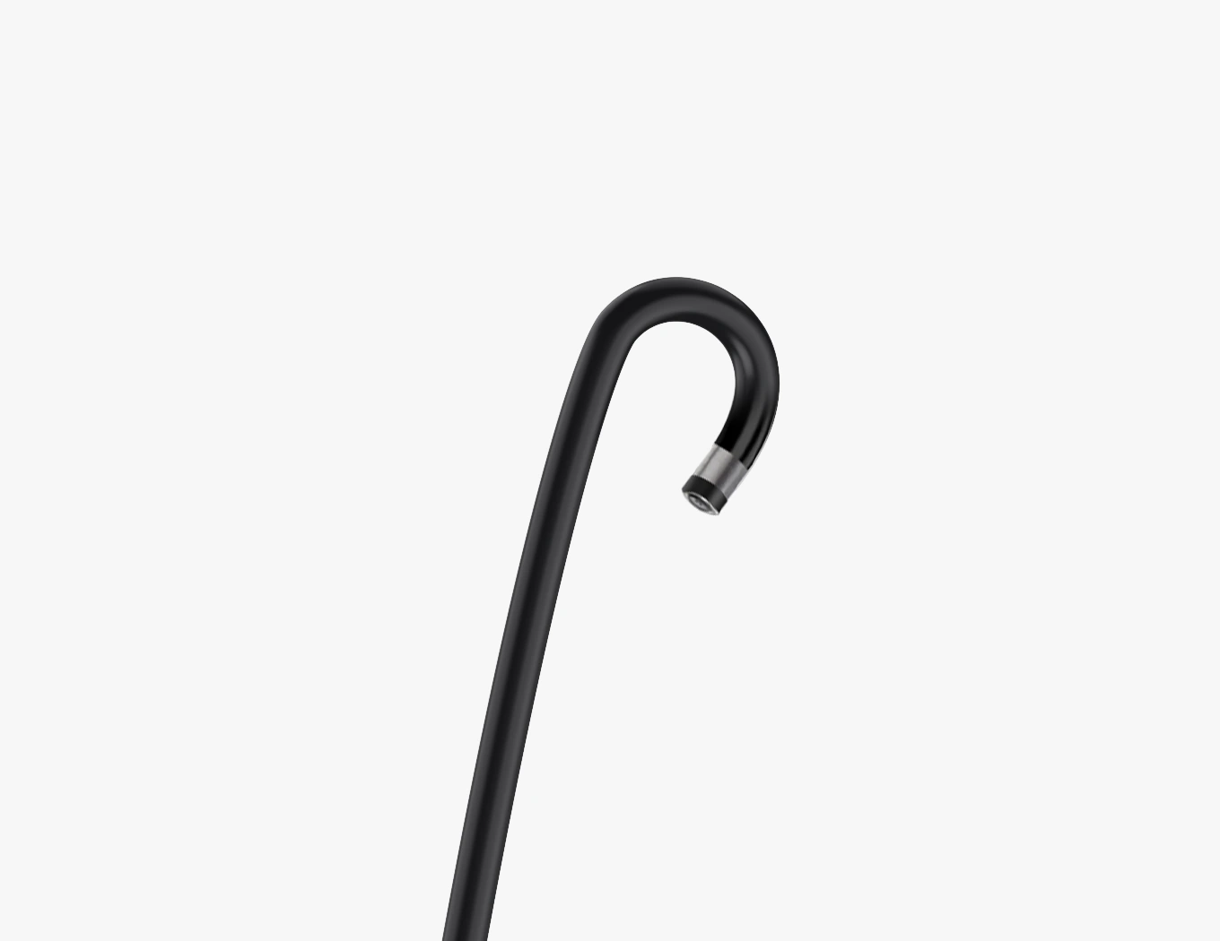
Flexible scopes provide tip control and are used for nasopharyngeal examinations. Their suitability depends on the required bend, access space and dimensions. A scope suitable for an oral or nasopharyngeal view is not automatically suitable for passage through a narrow nasal cavity. 4 5
Similarly, a setup selected for a larger dog should not simply be transferred to a toy-breed dog or cat. Smaller patients may require smaller equipment and a different access plan. 1
Choosing the scope and supporting equipment
Dimensions and access
Review insertion-tube diameter, maximum insertion-section width, distal-tip dimensions and working length. These measurements describe different parts of the scope. Where accessories add to the insertion profile, assess the complete configuration rather than selecting from a single diameter.
Viewing direction and maneuverability
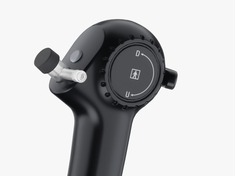
Match viewing direction and tip movement to the intended anatomy and route. Confirm the configuration’s suitability for the planned view; the name “rhinolaryngoscope” alone does not establish that it can perform every nasal or nasopharyngeal examination.
Instrument and channel compatibility
Decide whether instruments will pass through the scope or use another supported arrangement. For through-scope use, confirm channel size, instrument outer diameter and working length. A channel’s presence does not establish compatibility with every forceps, brush or retrieval accessory. 5
Observation-only models should not be described as providing through-scope instrument access. Conversely, a channel specification alone is not confirmation of a complete irrigation, suction or sample-collection setup.
Display, documentation and care
Check the required display, software and connections, including how still images or recordings are saved and transferred. Confirm which functions belong to the scope, host or software.
For reusable components, review the current manufacturer-specific cleaning, reprocessing, drying and storage instructions. Handle single-use components according to their labeling; do not apply a generic reusable-endoscope routine to every item. 5
LumiVET’s Veterinary Flexible Rhinolaryngoscope page separates observation-only and instrument-channel models. Compare the selected model’s full specifications with the intended access, instruments and display setup. Do not assume that all configurations support the same examination or sampling tasks.
Keeping the examination record useful
Plan how images, observations, sample locations and laboratory reports will be associated in the clinical record. For equipment evaluation, check image export and transfer, shared viewing and the software needed to retrieve captured media.
Recording capability supports this workflow; it does not replace written findings or interpretation. A distributor or equipment supplier can help clarify system functions, while clinical conclusions remain with the veterinary team.
Frequently asked questions
Does dog rhinoscopy replace a CT scan?
No. Endoscopy examines accessible surfaces and can support sampling; CT evaluates structures and disease extent that may not be visible endoscopically. The tests are selected and interpreted as parts of the patient’s investigation. 2
Can every flexible rhinolaryngoscope perform posterior rhinoscopy?
Suitability must be checked for the specific model and intended approach. Review dimensions, bending characteristics, required access space and intended use rather than relying on the product name or one angulation figure.
Does a working channel mean biopsy equipment is included?
No. Channel dimensions describe an access feature. The compatible instruments, required accessories and supplied configuration must be confirmed separately.
Review the configuration for your practice
Compare the Veterinary Flexible Rhinolaryngoscope models, then contact LumiVET with the patient range, intended examination, sampling needs and existing display equipment. For broader practice planning, explore the small-animal endoscopy options.
This overview is intended for veterinary professionals. It does not replace patient-specific clinical judgment, practical training or the instructions for the selected equipment.
References
- VCA Veterinary Care Animal Hospital and Referral Center. Rhinoscopy. Specialty-service overview.
- Rösch S, Oechtering GU. Insights into Canine Idiopathic Rhinitis from a Prospective Study: A Multimodal Diagnostic Perspective. Animals. 2026;16(10):1438. DOI: 10.3390/ani16101438.
- Lent SE, Hawkins EC. Evaluation of rhinoscopy and rhinoscopy-assisted mucosal biopsy in diagnosis of nasal disease in dogs: 119 cases (1985–1989). Journal of the American Veterinary Medical Association. 1992;201(9):1425–1429.
- McKiernan BC. Respiratory Endoscopy—A Visual Assessment of the Respiratory Tract. WSAVA 2001 congress proceedings; original lecture text available in a reproduced PDF.
- Callahan Clark J. Endoscopic Equipment for the Veterinary Practitioner. Today’s Veterinary Practice. November/December 2012.